Microvascular ischemic disease (MVID), also known as cerebral small vessel disease, is a brain condition predominantly affecting older individuals, with the risk increasing significantly with age. In a 2019 review, it was found that MVID affects about 5% of individuals aged 50, while the prevalence approaches nearly 100% for those aged 90 and above [1]. MVID is often associated with cognitive decline and can lead to severe conditions such as dementia and stroke if left untreated [2]. MRI plays a crucial role in diagnosing MVID. It enables the visualization of white matter hyperintensities (WMH), which are often indicative of arteriolar disease, a main component of MVID [3].
One unique aspect of MVID is its classification based on the extent of white matter hyperintensities (WMH) seen in MRI scans, often using the Fazekas scale. In a study involving 849 subjects aged between 21 to 79, individuals were categorized based on the 4-stage Fazekas scale according to the hyperintense lesions observed on T2-weighted FLAIR MRIs [4]. The Fazekas score is a reliable tool for evaluating the severity of MVID, with a higher score indicating more severe disease.
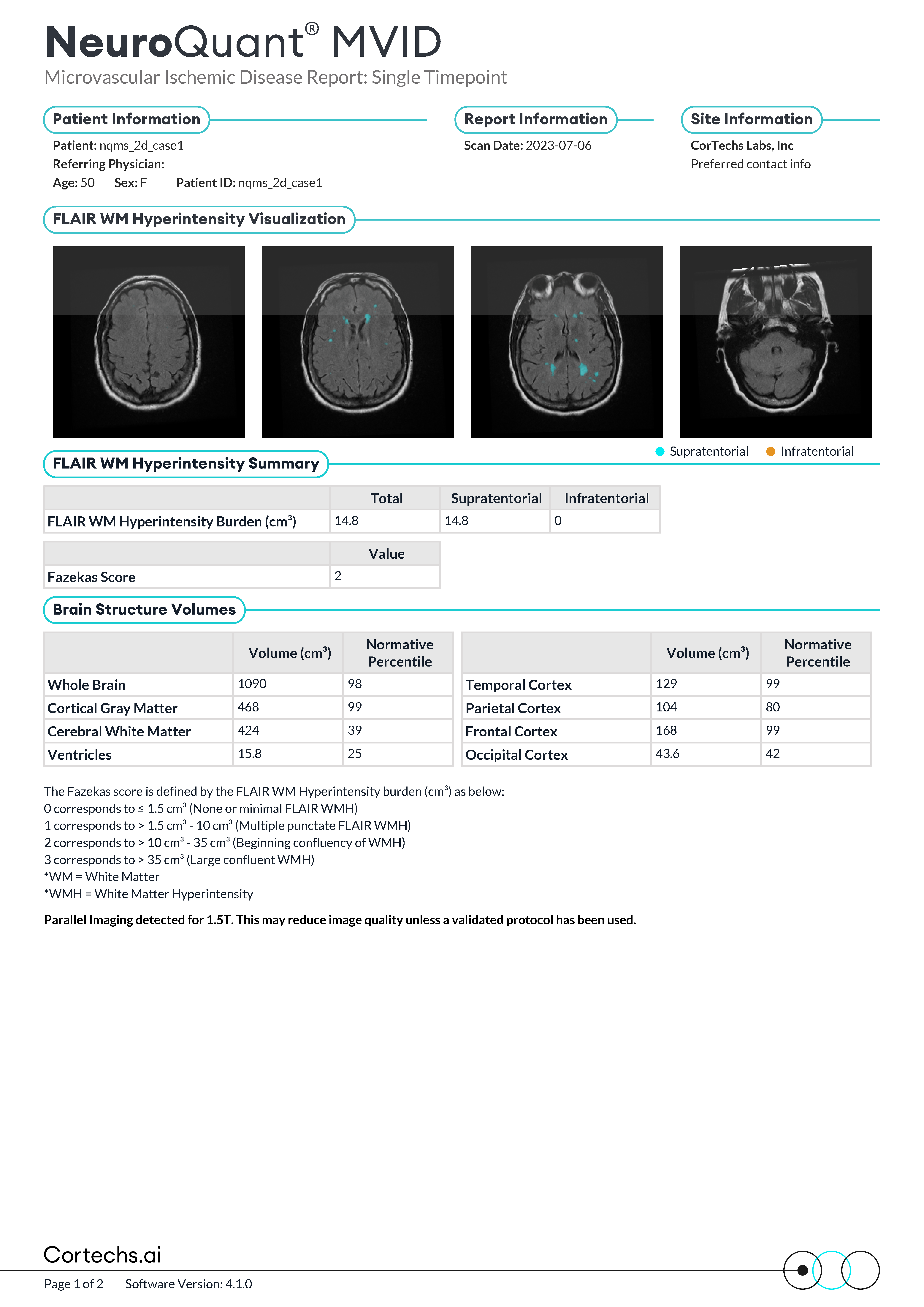
Our new microvascular report introduces a slight deviation from the traditional Fazekas scale. Instead of using two separate scales for Periventricular White Matter (PWM) and Deep White Matter (DWM), we have consolidated these into a single Fazekas score that assesses the total hyperintensity volume burden. This approach simplifies the assessment process and provides a more comprehensive overview of the disease burden. By evaluating the total hyperintensity volume, we can offer a clearer and more unified picture of a patient’s condition. This consolidation enhances the accuracy of diagnosis and monitoring, aiding in better clinical decision-making and treatment planning.
Two independent expert neuroradiologists applied a single Fazekas score (ranging from 0-3) to 77 FLAIR images from a cohort of normal patients. The T1 and FLAIR input series from these 77 cases were processed with NQMS 4.0 and, as part of the output, whole brain white matter hyperintensity load (mL) was calculated. Left-exclusive cutoff ranges in mL were determined for each Fazekas score, maximizing the average accuracy (0.90) as compared to the two physician raters.


Using the whole brain lesion volume as the thresholding feature, the following cutoffs were calculated using 65 cases of the dataset to provide the highest accuracy (>90%) on both independent expert ratings. The calculated thresholds were incorporated in NeuroQuant v4.1:
| Fazekas Score | Lower limit (mL): Greater than | Upper Limit (mL): Less than or equal to |
| 0 | 0.0 | 1.5 |
| 1 | 1.5 | 10 |
| 2 | 10.0 | 35 |
| 3 | 35.0 | N/A |
This NeuroQuant®️ MVID report innovation not only aligns with the latest advancements in neuroimaging, but also ensures that both physicians and patients receive clear and actionable insights into the severity of MVID. This approach facilitates better communication between healthcare providers and patients, ultimately contributing to improved patient outcomes and more targeted therapeutic strategies. Check out the report below:

References:
- https://doi.org/10.1212/WNL.0000000000007
- https://doi.org/10.1136%2Fsvn-2016-000035
- https://doi.org/10.1161/STROKEAHA.120.030063
- Kynast J, Lampe L, Luck T, Frisch S, Arelin K, Hoffmann KT, Loeffler M, Riedel-Heller SG, Villringer A, Schroeter ML. White matter hyperintensities associated with small vessel disease impair social cognition beside attention and memory. J Cereb Blood Flow Metab. 2018 Jun;38(6):996-1009. doi: 10.1177/0271678X17719380. Epub 2017 Jul 7. PMID: 28685621; PMCID: PMC5999004.


