COVID-19 Neurological Manifestations
Cortechs.ai is bringing the most widely used clinical brain morphometry tool to help with the COVID-19 pandemic. While COVID-19 primarily affects the lungs, the latest research shows that patients, especially ones with more severe infection, may also experience COVID-19 neurological manifestations of both within the central and peripheral nervous system, and the skeletal muscular symptoms.
Research shows that patients can present with COVID-19 neurological manifestations such as:
- Large vessel strokes, especially patients under the age of 50
- Encephalopathy in the acute setting and during hospitalization
- Swelling in the whole brain, and cortical brain regions, particularly the frontal and temporal lobes in some severe cases.
- Ataxia
- Anosmia
LesionQuant FLAIR Lesion Report, NeuroQuant Triage Brain Atrophy (TBA), and Custom COVID-19 reports will be available, free of charge, to all facilities for COVID-19 patients or for COVID-19 research. Below we will review a few COVID-19 case studies using NeuroQuant and LesionQuant for volumetric MRI analysis.
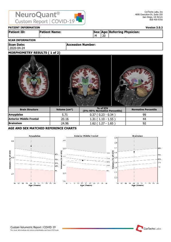
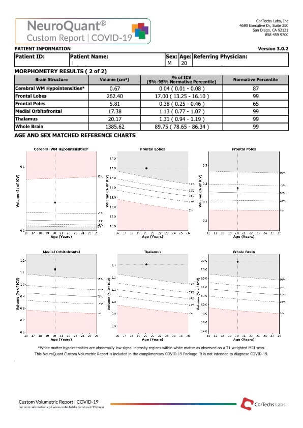
Case 1: Custom COVID-19 Report
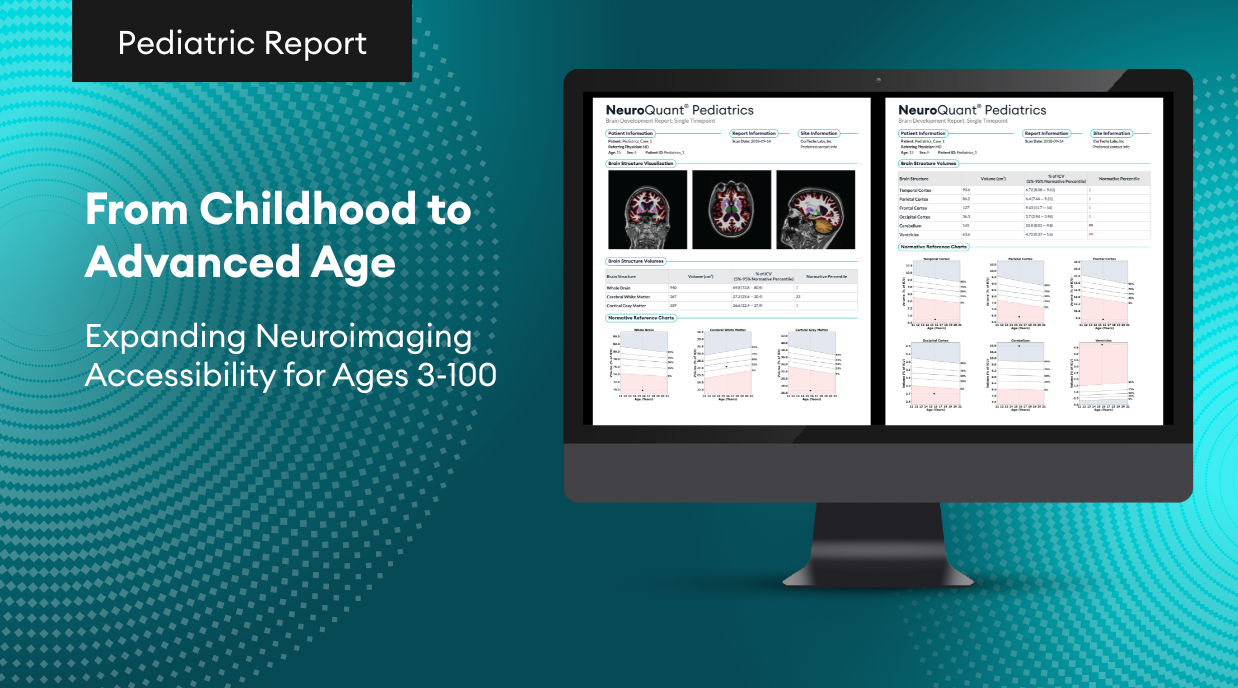
The Custom COVID-19 Report includes brain structures that are potentially affected by COVID-19: cerebellum, brainstem, whole brain, frontal lobes, frontal poles, medial orbitofrontal lobes, anterior middle frontal lobes, thalamus, amygdalae, and cerebral white matter hypointensities. This report will show the raw volume, percent of ICV, and normative percentiles for each structure. NeuroQuant’s Custom volumetric reports were designed to provide users with flexibility in report design. The predefined COVID-19 custom report is an example of how users can create their own NeuroQuant report with structures that meet their specific needs.
Structures Affected (based on recent research)
- Amygdalae (99th)
- Anterior Middle Frontal
- Brainstem (92nd)
- Cerebral WM Hypointensities (87th)
- Frontal Lobes (99th)
- Frontal Poles
- Medial Orbitofrontal (99th)
- Thalami (99th)
- Whole Brian (99th)
Patient History:
This 20-year-old male patient was treated for COVID-19 in early February. He tested positive but was not admitted to the hospital. Roughly two months later he returned presenting neurological manifestations; confusion, dizziness, visual impairment, ataxia, and seizures. A brain MRI was ordered with NeuroQuant analysis using both the Custom and TBA reports.
NeuroQuant Findings:
Findings demonstrate statistically significant enlargement of the amygdalae in the 99th percentile, the brainstem in the 92nd percentile, frontal lobes in the 99th percentile, the medial orbitofrontal region in the 99th percentile, the thalami in the 99th percentile, and the whole-brain in the 99th percentile. Cerebral white matter hypointensities are borderline high in the 87th percentile.
Impression:
Volumetric analysis demonstrated statistically significant enlargement of the amygdalae, brainstem, frontal lobes, medial orbitofrontal region, thalami, and whole brain. Cerebral white matter hypointensities are in the
higher range, but not clearly statistically significant.
Case 2: Triage Brain Atrophy Report
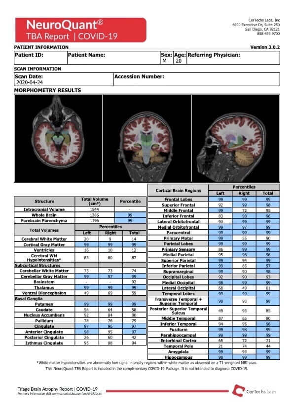
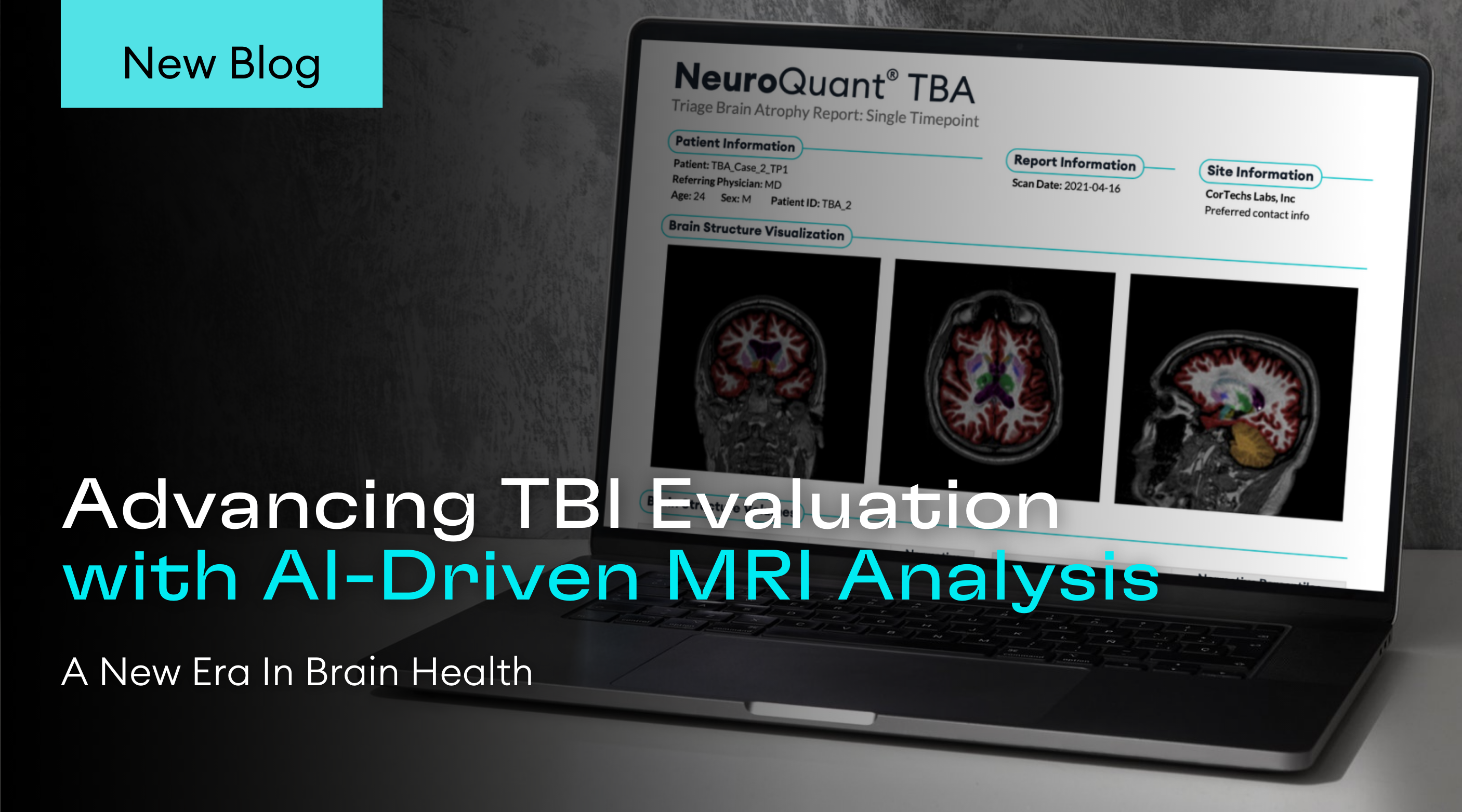
The TBA report is included so clinicians can quickly identify swelling in the whole brain, and brain regions, particularly the frontal and temporal lobes as seen in some severe COVID-19 cases. For encephalitis, clinicians can quickly review volumes for the whole brain, individual lobes, and numerous structures relevant to COVID-19 neurological manifestations, providing an immediate overview from both a global and individual structure perspective.
Patient History:
This is the same 20-year-old male patient that was treated for COVID-19 in early February and presented with confusion, dizziness, visual impairment, ataxia, and seizures two months following his diagnosis. A brain MRI was ordered with NeuroQuant analysis using both the Custom and TBA reports.
NeuroQuant Findings:
Within this TBA report, there are numerous clusters of blue indicating global edema. Specifically, we see the whole-brain and forebrain parenchyma in the 99th & 98th percentiles respectively. Next, we see cortical gray matter, cerebellar gray matter, and cingulate above the 95th percentiles. There is also statistically significant multifocal edema, notably involving the frontal, and parietal lobes bilaterally, and the occipital and temporal lobes with a left temporal predilection.
Impression:
The volumetric analysis demonstrates statistically significant multifocal edema, notably involving the frontal, and parietal lobes bilaterally, and the occipital and temporal lobes with a slight left temporal predilection. This report provided an objective, quantifiable explanation for his neurologic manifestations.
Case 3: LesionQuant FLAIR Lesion Report (Two-Timepoints)
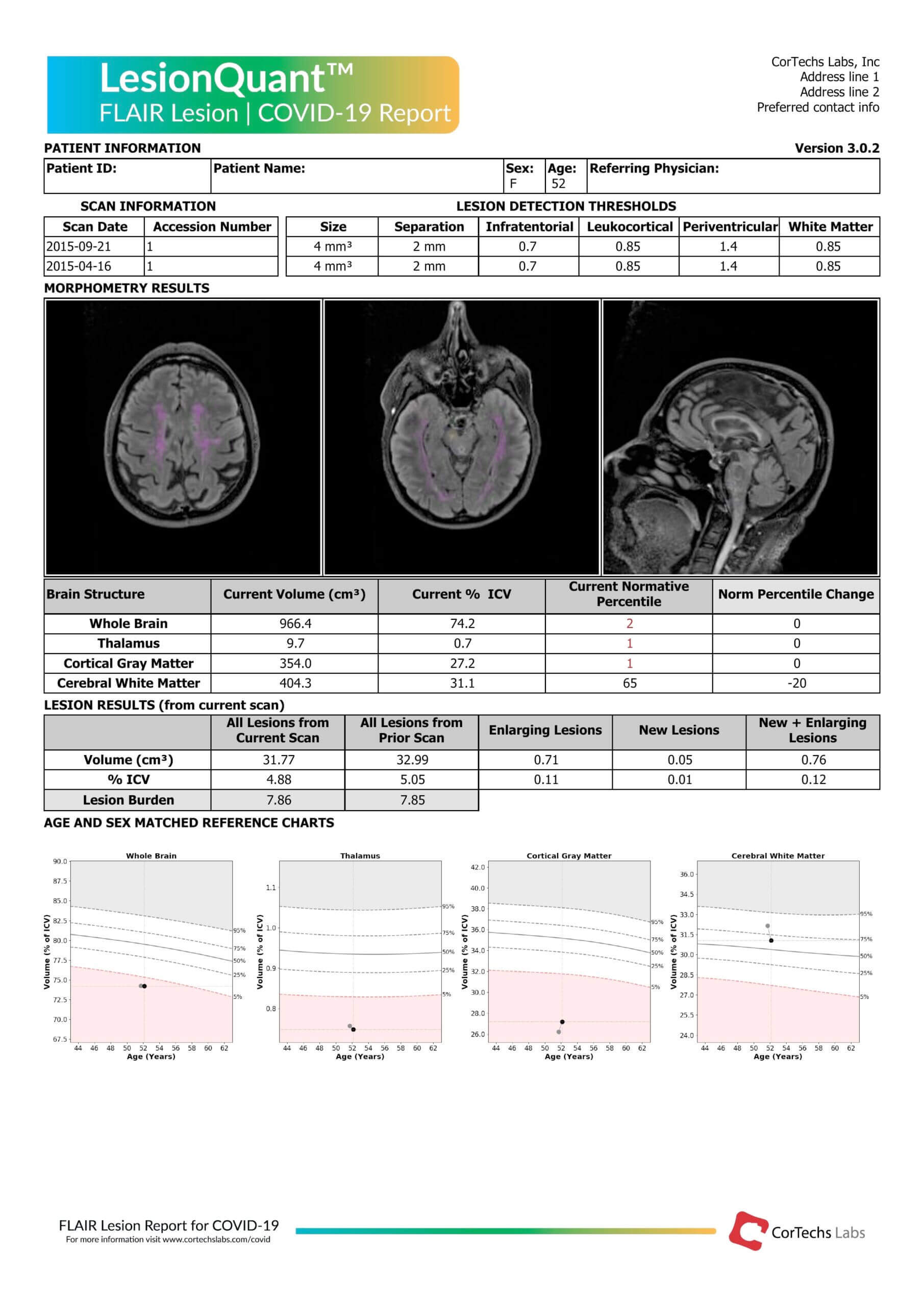
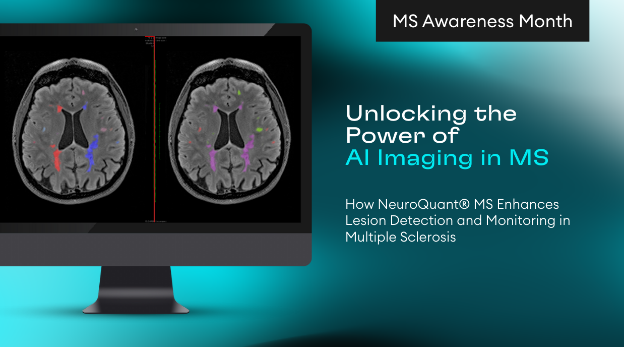
According to recent clinical findings, COVID-19 patients under the age of 50 are often presenting with large vessel strokes. The LesionQuant report has the unique feature of giving detailed volumetric structural brain measurements in addition to lesion volumes, percent of intracranial volume, and total lesion burden.
With speed and accuracy, LesionQuant performs automated FLAIR lesion segmentation and quantification along with the following brain regions quantification: whole brain, thalamus, cortical gray matter, and cerebral white matter. LesionQuant analysis may be useful in evaluating a brain with micro strokes and for short-term care because it can guide more aggressive anticoagulant or antiplatelet therapy. It may also be useful for long-term longitudinal tracking to measure drug effectiveness and to determine whether changes in disease-modifying therapies need to be altered.
Patient History:
In this final case study, we have a 52-year-old female patent with known MS who was admitted to the ICU with COVID-19. She had worsening neurologic symptoms and underwent a brain MRI. This report compared the current scan to a scan acquired two months prior, before her COVID-19 diagnosis. You can see how volumetrics are useful in tracking the disease trajectory of this MS patient with COVID-19
NeuroQuant Results: Analysis found statistically significant whole-brain volume at the 2nd percentile, the thalami, and cortical gray matter in the 1st percentile. below the normative 5th-95th percentile range. The cerebral white matter falls within the normative range between the 5th-95th percentiles at the 65th percentile but has reduced in volume from the previous scan, contributing to a -20 percentile change.
Lesion Results:
- Total lesion volume – current scan 31.77 cm3
- Lesion percent of ICV is 4.88%
- Total lesion burden of 7.85
Impression:
LesionQuant analysis demonstrates evidence of demyelinating disease with a severe plaque burden (7.85). The volume of the total plaque burden is 31.77 cm3. Modestly progressive in degree since the prior exam (with 0.15% interval increase in plaque burden).
Request Access to the Covid-19 Report Package
The NeuroQuant and LesionQuant reports offered as part of the complementary COVID-19 package are to be used for follow-up of COVID-19 patients. They are not intended for COVID-19 diagnosis. LesionQuant’s FLAIR Lesion Report and NeuroQuant’s TBA and Custom Report are free for clinicians and researchers.
