Using NeuroQuant for volumetric imaging of patients with TLE.
Neuroimaging is one of several important tools physicians use to help provide objective disease assessment and effective treatment planning for patients with epilepsy. MRI volumetric analysis using sophisticated AI-based software has become a significant tool in the evaluation of patients with epilepsy and is especially helpful in assessing the possibility of surgery.
Volumetric MRI tools, such as NeuroQuant®, allow for the segmentation and measurement of brain structures to provide anatomic and structural information about the brain in a non-invasive fashion.

NeuroQuant is a leading medical device software that accurately segments brain structures and calculates each structure’s volume from 3D MRI images. It compares the volumes to a normative database comprised of individuals of the same age and sex. The use of volumetric measurements of temporal lobe structures can help physicians facilitate the recognition of subtle hippocampal atrophy, which may have not been identified by visual exam alone. Since the arrival of brain MR imaging, hippocampal atrophy is recognized as the most frequently seen neuroradiologic abnormality in temporal lobe epilepsy (TLE). Using NeuroQuant for volumetric imaging of patients with TLE provides physicians with an objective and precise measurement in their assessment of hippocampal atrophy.
Neuroimaging studies suggest that TLE is associated with pathologic changes in hippocampal physiology and morphology. A history of febrile seizures is associated with hippocampal volume loss in patients with TLE. Asymmetric hippocampal atrophy, beyond what is considered normal hippocampal asymmetry, may be a sign that the atrophied hippocampus could be the source of the epileptic seizures.
In patients with TLE, there can often be vague EEG findings and clinical symptoms that require an MRI to help determine the location of seizure onset. While it is possible for moderate to severe hippocampal atrophy to be detected by visual examination of the MRI scan, volumetrics can be particularly helpful when the asymmetry between the two hippocampi is less than 5-10 percentile. In a visual read, radiologists are looking for a difference in volumes of the hippocampi. However, sometimes the tilt of a patient’s head or abnormal shapes of the hippocampus can make it difficult to determine whether one hippocampus is significantly smaller than the other.
The volumetric results of an MRI scan are objective quantitative measurements that are reproducible and objective. This can help highlight cases where there is slight unilateral hippocampal or bilateral volume loss. A 2015 study, Mesial Temporal Sclerosis: Accuracy of NeuroQuant versus Neuroradiologist, found that NeuroQuant performed better than expert Johns Hopkins Hospital trained neuroradiologists, with NeuroQuant having a hippocampal asymmetry classification accuracy of 79.4% versus the neuroradiologists’ accuracy of 72.6%.
For physicians evaluating TLE, the NeuroQuant Hippocampal Asymmetry report provides volume measurements of the left and right hippocampus, the asymmetry between the left and right hippocampus, and it indicates the patient’s hippocampal volume measurements as compared to a healthy population via an extensive, proprietary normative database. This allows NeuroQuant to deliver a precise indication and visualization of where that individual’s hippocampal volumes and asymmetry lie within an age- and sex-matched reference chart. The NeuroQuant Hippocampal Asymmetry report delivers precise and critical information to aid physicians in their assessment of TLE.
Sample Case: 43-year-old woman with suspected temporal lobe epilepsy
Patient History: This patient was undergoing further evaluation for temporal lobe epilepsy. She could no longer work or drive her car and was potentially a surgical candidate for an amygdalohippocampectomy.
NeuroQuant Findings:
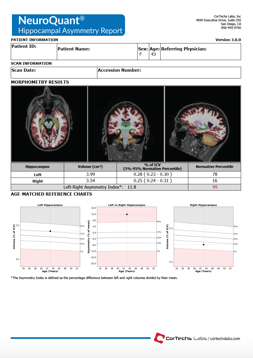 Left hippocampus is within normative 5th-95th percentile range for the same age and sex at the (3.99 cm3) 93rd percentile
- Right hippocampus in normal statistical limits at about one standard deviation below normal range for the same age and sex at the (3.54 cm3) 37th percentile
- Left to right asymmetry index is abnormal at the (11.8 cm3) 99th percentile
Impression: Together this is evidence of asymmetric hippocampi – a key data point in the assessment of TLE.
Treatment and Outcome: Independently, each hippocampal volume falls within the 5th-95th percentile range. However, when evaluating the asymmetry, it is apparent that the right hippocampus is significantly smaller than the left hippocampus, which confirmed the initial suspicion of right-sided TLE. This patient went on to have an amygdalohippocampectomy and fully recovered, allowing her to return to her career.


