Volumetric quantification of focal atrophy in AD

Fully-automated quantification of regional brain volumes for improved detection of focal atrophy in Alzheimer’s disease

Volumetric analysis of structural MR images of the brain may provide quantitative evidence of neurodegeneration and help identify patients at risk for rapid clinical deterioration. This note describes tests of a fully automated MR imaging postprocessing system for volumetric analysis of structures (such as the hippocampus) known to be affected in early Alzheimer disease (AD). The system yielded results that correlated highly with independent computer-aided manual segmentation and were sensitive to the anatomic atrophy characteristic of mild AD.

NeuroQuant® Results Published in the American Journal of Neuroradiology.

[button]Download[/button]
Insights

02/15/2025

NeuroQuant® Dementia: Advancing Alzheimer’s Disease Care in the Era of DMTs

AI-driven brain volumetric analysis is revolutionizing Alzheimer’s care, providing precise diagnostic support, monitoring, and early detection.

02/09/2025

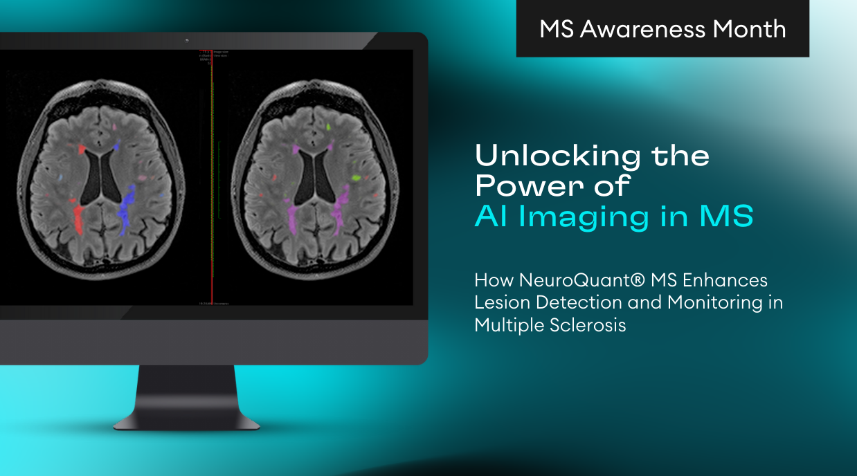
NeuroQuant® MS:  Advancing Precision in Multiple Sclerosis

Dr. Suzie Bash discusses NeuroQuant MS, noting its ability to segment plaques, analyze volume, and track lesion burden for assessing disease progression

02/09/2025

Epilepsy Care with NeuroQuant Seizure: The Power of Quantifying Hippocampal Asymmetry

Accurate imaging of the hippocampus is crucial for effective diagnosis and treatment planning in TLE, with structural abnormalities linked to seizure onset
Scroll to Top