By
Cortechs.ai
2 mins
Previously, we discussed the importance of image distortion correction and alignment to a standard space, providing the ability to measure accurately and repeatedly and track brain volumes over time. Additionally, we examined how Cortechs.ai’ products deliver high image quality standardization through our automatic atlas-based registration and spatial distortion correction techniques.
While each modality (MRI, PET, CT) has its own set of uncertainties and image artifacts, Cortechs.ai’ image distortion correction, and image consistency and reproducibility technologies eliminate a majority of the alignment problems. Additionally, Cortechs.ai’ unique, patented Dynamic Atlas™ alignment technology creates a joint coordinate system that allows image data sets from each modality to reference, facilitating, for example, MRI + PET image overlay.
PET Brain Studies Powered by NeuroQuant
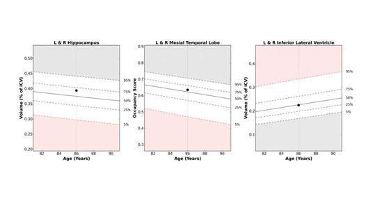
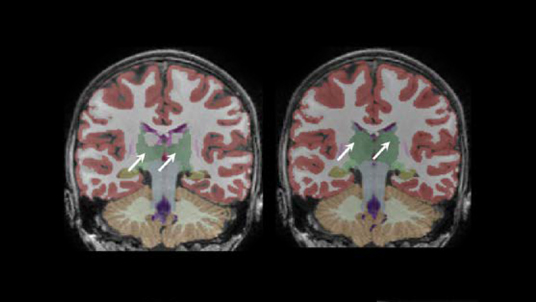
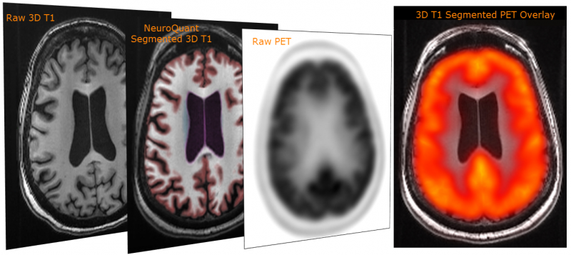
PETQuant™ offers clinicians the ability to quantify subregional tracer binding in native brain space. How does it do this? Rather than providing deposition of amyloids in a template volume that might or might not correlate to the actual patient’s frontal lobe or hippocampus, NeuroQuant® provides deposition information (and norms) in volumes that are derived directly from the patient’s 3D T1 images precisely indicating relevant brain structure information for the clinician.
The patient’s native space provides the actual and accurate deposition information – not just approximates in general volumes. The patient’s native space becomes even more important when evaluating brain structure changes over time. Taking the patient’s brain structure volume changes into account allows for a more accurate amyloid deposition measurement because the associated reference volumes change appropriately.
The same principle can be applied to other modalities, such as comparing 3D T1 to 3D T1 for measuring changes and following a patient longitudinally, analysis of 3D T1 to T2 FLAIR for lesion segmentation and disease progression evaluation, as well as evaluating other MRI and non-MRI imaging modalities.
![]()
Share