By
Cortechs.ai
2 mins
Cortechs.ai is providing the most widely used clinical brain morphometry tool to help with the follow-up of COVID-19 patients and to contribute to neurological research of the pandemic.
As the COVID-19 pandemic progresses, there are reports from different institutions around the world of neurological symptoms and manifestations in the central and peripheral nervous systems (CNS, PNS). Due to these findings, Cortechs.ai is offering a new complimentary brain volumetric report package.
Our LesionQuant FLAIR Lesion Report, NeuroQuant Triage Brain Atrophy (TBA) and Custom COVID-19 reports are available, free of charge, to all facilities for COVID-19 patients or for COVID-19 research. Through the use of our automated software, it is possible to monitor the volumes of 75 brain structures and regions, as well as any changes in lesion burden or lesion distribution over time. As the research on the neurological effects of the disease is ongoing, our offering may be updated accordingly.
While COVID-19 primarily affects the lungs, the latest research (below) shows that patients, especially ones with more severe infection, may also manifest neurologic symptoms of the CNS, PNS, and skeletal muscular symptoms. Cortechs.ai’ existing fully automated brain volumetric solutions can be easily customized to provide COVID-19 relevant brain volumetric information in minutes.
COVID-19 patients may manifest neurologic symptoms, which, according to Mao et al. (2020) in an article published in JAMA Neurology [3], are more likely to manifest in patients with more severe infection. In this study, 36.4% of patients experienced:
CNS Symptoms:
- Dizziness
- Headache
- Impaired consciousness
- Acute cerebrovascular disease
- Ataxia
- Seizures
PNS Symptoms:
- Taste impairment
- Smell impairment
- Vision impairment
- Nerve pain
Mao and colleagues found that some patients develop COVID-19-related symptoms only after showing neurologic symptoms, but the rate of neurological symptoms is higher in patients with more severe respiratory disease status1. According to Kandemirli et al. (2020) in a study published by the Radiological Society of North America [4], of those patients in the intensive care unit with COVID-19 infection presenting neurologic symptoms who underwent MRI, 44% (12/27) had abnormal MRI findings.
While Cortechs.ai’ NeuroQuant and LesionQuant volumetric software were not designed to diagnose COVID-19, they can provide longitudinal quantitative data to inform clinicians about brain regions the disease could be affecting and automatically compare the results with others of the same age and sex. This may help physicians detect and address any findings that are treatable.

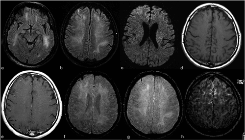
The MRI images here demonstrate an exam of a 59-year old intubated male patient with altered mental status. The FLAIR images demonstrate prominent symmetric white matter hyperintensity and right frontal cortical hyperintensity. There is also linear hyperintensity within the frontal sulci. These hyperintensities will be automatically quantified and tracked longitudinally with LesionQuant.
Complimentary COVID-19 Report Package
The LesionQuant FLAIR Lesion Report, NeuroQuant Triage Brain Atrophy (TBA) and Custom COVID-19 reports are available in a complimentary package. These reports segment and quantify patient brain structures and lesions in under 30 minutes. As the research on the neurological effects of the disease is ongoing, our offering may be updated accordingly.
This complimentary report package is intended to be used in the follow-up of patients who have been diagnosed with COVID-19 and are experiencing neurological symptoms.
Apply for free access to the NeuroQuant and LesionQuant COVID-19 report package
Interested in learning more about how NeuroQuant and LesionQuant can be used to help assess possible neurological manifestations of COVID-19?
References
- WHO Timeline – COVID-19. (2020). Retrieved 28 May 2020, from https://www.who.int/news-room/detail/27-04-2020-who-timeline—covid-19
- Li YC, Bai WZ, Hashikawa T. The neuroinvasive potential of SARS-CoV2 may play a role in the respiratory failure of COVID-19 patients. J Med Virol. February 27 2020:10.1002/jmv.25728. doi: 10.1002/jmv.25728. Epub ahead of print.
- Mao L, Jin H, Wang M, et al. Neurologic Manifestations of Hospitalized Patients With Coronavirus Disease 2019 in Wuhan, China. JAMA Neurol. Published online April 10, 2020. doi:10.1001/jamaneurol.2020.1127
- Kandemirli, Sedat G et al. “Brain MRI Findings in Patients in the Intensive Care Unit with COVID-19 Infection.” Radiology, 201697. May 8, 2020, doi:10.1148/radiol.2020201697
Share


