Continuing our educational series on using volumetric brain imaging in the assessment of neurodegenerative conditions, the Cortechs.ai team welcomed a board-certified, internationally recognized neurologist dedicated to improving diagnosis and care for people with multiple sclerosis (MS) and related disorders to our San Diego home office. The thought-provoking presentation focused on diagnosing multiple sclerosis, treatment options and the use of volumetric MRIs for prognosis and drug treatment decisions.
MS is a devastating and costly chronic disease and while the symptoms can be treatable in many cases, there is currently no cure. Affecting roughly 2 million people worldwide, most people are diagnosed with MS between the ages of 20 and 50 and more people are being diagnosed with MS today than ever before. While the reason for this increase is unknown, likely possibilities include greater disease awareness, increased availability of medical care and improved diagnostic capabilities. Currently in the US, MS diagnoses and treatments total 1% of the total US healthcare budget (including medical costs, personal care and assistance) with MS drugs alone averaging $60,000 per patient per year.
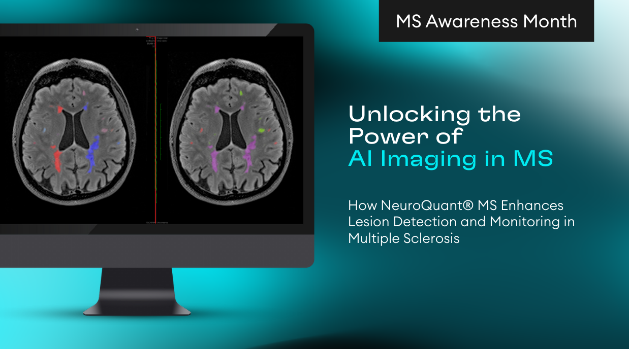
Diagnosing multiple sclerosis is a waiting game. There is not a single test to make a diagnosis, symptoms vary from patient to patient, making it necessary to rule out other conditions and explanations. In people with MS, cognitive slowing is observed in the ability to process tasks, as well as planning and sequencing activities. When an MS diagnosis is made, a physician has found evidence of two episodes of disease activity (lesions) occurring in the central nervous system at different points in time. These lesions are usually found in the gray matter, where muscle control and activity processing occur.
Magnetic Resonance Imaging (MRI) is used in the diagnosis and is an integral part in the management of MS. A MRI is taken to finalize a MS diagnosis by providing evidence of the new and enhancing lesions. After MS is confirmed, drug treatments are started to prevent relapse and a baseline MRI scan is taken. Repeating MRI scans are taken periodically (6, 18 and 36 months) after treatment has started, to ensure that it continues to be effective.
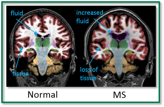
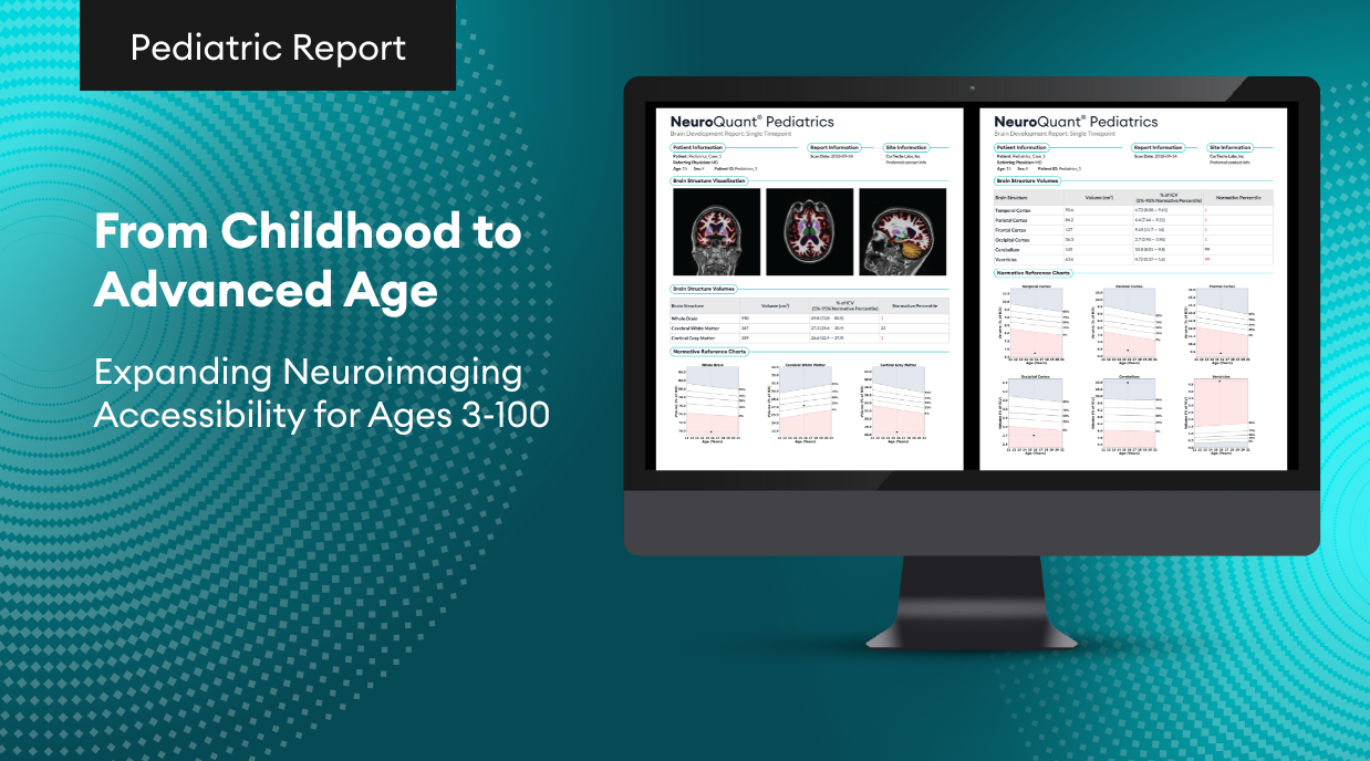
NeuroQuant® is a leading medical device software for volumetric MRI processing, providing automatic image segmentation of brain structures. NeuroQuant uses the images from the MRI scan to measure the volume of certain brain structures. In this case, volume measurements of the whole brain, lateral ventricles, thalamus, gray matter, white mater, 3rd ventricles, hippocampus, inferior lateral ventricles and T1 white matter hypointensities are reported, providing important information in the assessment of multiple sclerosis. For MS, the NeuroQuant Multi Structure Atrophy Report can be used to help assess volume changes in several brain structures during an initial measurement and follow up measurements to help physicians monitor MS and treatment effectiveness. This longitudinal feature allows physicians to quickly and easily compare brain structure volume changes and rate of change at multiple scan intervals, on the same report.
- De Stefano, Nicola et al. (2014). Clinical Relevance of Brain Volume Measures in Multiple Sclerosis. CNS Drugs 28:147–156
- Fisher, Elizabeth et al. (2008). Gray Matter Atrophy in Multiple Sclerosis: A Longitudinal Study. Ann Neurol. 64:255–265
- The Global Impact of Multiple Sclerosis. May 2010. Prepared for Multiple Sclerosis International Federation, London, United Kingdom, by RTI International.
- http://www.nationalmssociety.org/
