Did you know? NeuroQuant Provides Color-Coded Segmentations for Easy Structure Review

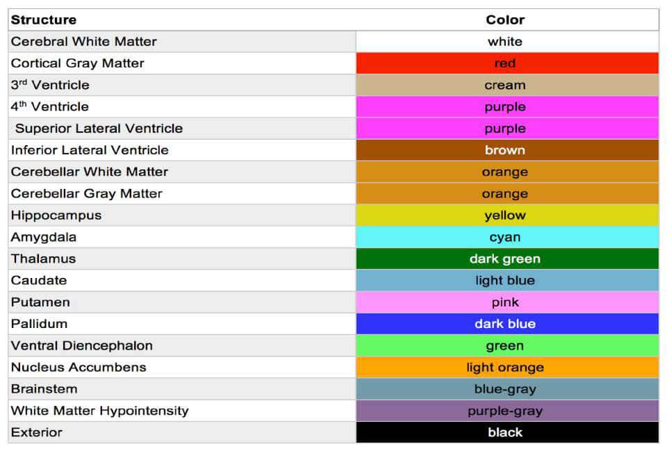
In addition to providing automated and fast brain structure segmentation, NeuroQuant produces color-coded overlays in which each color represents a different region of interest. The color-coding allows physicians to effortlessly review the segmentation. Also, for physicians wanting to examine a structure or structures that are not on the NeuroQuant output report, they can easily locate the structure by its associated color.

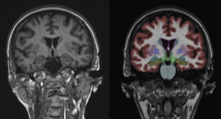
The images below show an MR T1-weighted images (left) and NeuroQuant segmentation overlaying a T1-weighted image (right). The table below the images provides the structure and associated color.

Cortechs.ai | Did you know? NeuroQuant Provides Color-Coded Segmentations for Easy Structure Review

Segmented Color-Blended Volumes Cortechs.ai | Did you know? NeuroQuant Provides Color-Coded Segmentations for Easy Structure Review

To learn more about the importance of using quantitative volumetric imaging for brain MRI in clinical practice, check out our latest Cortechs.ai white paper.

To see how you can use automated quantitative analysis to more objectively analyze your patients’ data, visit our website.
Insights

02/06/2025

Cortechs.ai and EDAP Announce World’s First OnQ™ Prostate-Assisted Focal One® Robotic HIFU Procedure at UCSF Prostate Cancer Center

This milestone marks the first clinical use of OnQ Prostate’s RSI technology to guide focal therapy, improving lesion localization and targeting for urologists.

02/02/2025

NeuroQuant® Brain Tumor: Revolutionizing Neuro-Oncology with AI-Driven Precision

Dr. Suzie Bash and Dr. Jeff Rudie dive deep into the advanced functionalities and clinical benefits of the tool that is revolutionizing neuro-oncology

01/28/2025

Revolutionizing Imaging Diagnostics with Cortechs.ai CEO, Kyle Frye

Hear from CEO Kyle Frye on the SCALE Community Podcast about how Cortechs.ai is transforming healthcare with cutting-edge AI-powered medical imaging solutions.
Scroll to Top