NeuroQuant MS sets a new standard in the assessment of multiple sclerosis (MS) on brain MRI, leveraging advanced AI-powered deep learning technology to empower clinicians with precise and efficient quantitative evaluation of brain changes associated with the disease.
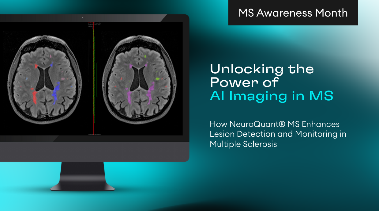
Dr. Suzie Bash, MD, a Medical Director at RadNet and Chief Medical Officer at Cortechs.ai, spoke with Applied Radiology at RSNA 2024, highlighting the capabilities of NeuroQuant MS. “The software color segments plaques by region, then performs automated volumetric analysis of both intracranial plaque burden and brain substructures. It also evaluates dynamic changes in lesion burden over time, offering essential insights into disease progression,” she explains.
“NeuroQuant MS has significant clinical utility since plaque volume, cortical gray matter volume, and whole brain volume are well-established prognostic biomarkers in multiple sclerosis,” states Dr. Bash.
NeuroQuant MS Optimizes Treatment
With approximately 20 disease-modifying therapies (DMTs) available for the treatment of multiple sclerosis (MS), precise evaluation of dynamic changes in plaque burden through serial annual MRIs is essential for informing therapeutic decisions.
“The unparalleled value of NeuroQuant MS lies in its ability to accurately monitor disease progression, facilitating the development of personalized, optimized treatment strategies that enhance patient outcomes,” explains Dr. Bash. “Neurologists rely heavily on neuroradiologists to provide accurate image interpretation since it directly influences decisions regarding whether a DMT should be substituted to improve efficacy,” she notes.
Challenges in Monitoring MS Plaque Burden
While the evaluation of plaque burden presents minimal challenges in patients with few plaques, MS is a chronic progressive disease, and many patients develop moderate to severe plaque burden over time. In such cases, accurately identifying new, enlarging, or shrinking plaques can be difficult, especially when considering differences in slice sampling, head angulation, field strength, and vendor variability between imaging time points.
Advancing Accuracy with Automation
Dr. Bash emphasizes that computers excel at pattern recognition compared to human observation, making automated solutions highly effective in overcoming the challenges associated with visual analysis of high disease burden states. The use of automated analysis significantly improves diagnostic accuracy, with studies reporting a 52% increase in the detection of MS disease activity using automated assessment compared to subjective visual interpretation.
Automated analysis also significantly enhances intra-reader and inter-reader consistency in lesion counts. Furthermore, quantitative assessment provides referring physicians with more valuable information than qualitative readings, as precise, objective measurements of plaque burden can be serially tracked across multiple examinations. This capability enables the identification of treatment efficacy trends over time, offering critical insights into disease progression and therapeutic impact.
Improved Efficiency in MS Imaging Interpretation
With increasing imaging volumes each year, radiology departments face challenges such as the clinical and financial impact of delayed report turnaround times. Radiologists also experience mounting pressure to maximize productivity to compensate for these upward volume trends, particularly with compensation increasingly tied to relative value units (RVUs). These demands place significant strain on radiologists to interpret images more rapidly without compromising accuracy.
“NeuroQuant MS addresses efficiency challenges by automating plaque burden report outputs, with essential findings included in a pre-populated report summary. This summary can be seamlessly integrated into dictation systems, such as Power Scribe, streamlining workflow and saving time,” notes Dr. Bash.
Studies indicate that the utilization of quantitative post-processing software with pre-populated reporting templates allow neuroradiologists to interpret 38% more MS cases per hour compared to subjective visual analysis. This improvement highlights the potential of automation to enhance workflow efficiency while maintaining diagnostic accuracy in the context of increasing imaging demands.
Clinical Feedback
NeuroQuant MS report outputs have garnered strong support from both neurologists and neuroradiologists.
“Data is at the core of everything we do,” says Kyle Frye, CEO of Cortechs.ai. “The longitudinal capabilities of our technology have received exceptional feedback, with referring physicians expressing confidence in the accuracy and reliability of the data they receive.”
Dr. Bash emphasizes that her referring clinicians consistently rely on NeuroQuant MS for the clinical assessment of their MS patients, recognizing it an essential tool. “My referrers agree that this state-of-the-art solution redefines what’s possible in MS care, with highly accurate and efficient quantitative insights informing optimal therapeutic management and improved quality of life for patients worldwide,” she notes. The practicality of NeuroQuant MS’ cloud-based seamless workflows provides neuroradiologists with efficient, detailed report outputs in under 6 minutes, enhancing workflow and productivity.
NeuroQuant MS represents a major advancement in precision medicine, empowering clinicians to streamline radiology workflows, enhance diagnostic accuracy, maximize efficiency, while optimizing MS patient care.


