Predicting Progression of Mild Cognitive Impairment to Alzheimer’s Disease: Hippocampal Occupancy Score (HOC)
The NeuroQuant Dementia report includes a Hippocampal Occupancy (HOC) Score and volumes of the hippocampi, superior and inferior lateral ventricles, and the entorhinal cortex, which are important biomarkers used in predicting progression of Mild Cognitive Impairment (MCI) to Alzheimer’s disease (AD). As with all NeuroQuant reports, the brain structure volumes and the HOC are compared to healthy patients of the same age and sex, normalized for intracranial volume (ICV).
Current research has demonstrated the effectiveness of the HOC in assessing patient progression from mild cognitive impairment (MCI) to Alzheimer’s disease (AD). This proven biomarker is an invaluable factor in predicting the progression of neurodegenerative diseases as approximately 10 – 15% of all patients diagnosed with MCI will develop AD annually [1]. Hippocampal volume remains the best single predictor of conversion of MCI to AD at a 3-year follow-up [2].
The HOC measures the degree of hippocampal atrophy accounting for volume loss and compensatory inferior lateral ventricle (ILV) expansion. It is calculated as a ratio of hippocampal volume to the sum of the hippocampal and ILV volumes in each hemisphere separately, which are then averaged and normalized for age and sex. The equation is as follows:

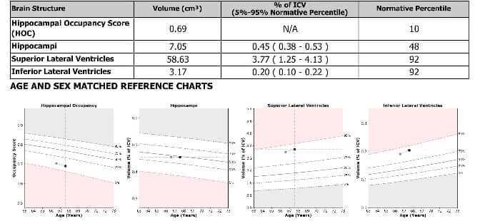
The NeuroQuant Dementia report provides the HOC in the first row of the table below the segmented images. The HOC is then visualized on the graph below the table to the far left, where it is plotted against the age and sex normalized values. The normative range (5th – 95th percentile) is represented in the white area of the graph while the solid line represents the mean.
Below are a few examples to better illustrate how the HOC is interpreted with the rest of the information provided on the NeuroQuant Dementia report.
Example Cases
Case 1:

- Two T1 MRI scans of a 67-year-old male patient.
- HOC measures .69 cm3 which is within two standard deviations of the normative range for age and sex.
- The hippocampi, lateral ventricles, and inferior lateral ventricles are all within two standard deviations of the normative range for age and sex.
- Conclusion: Normal follow-up that does not support neurodegeneration or the progression from MCI to AD.
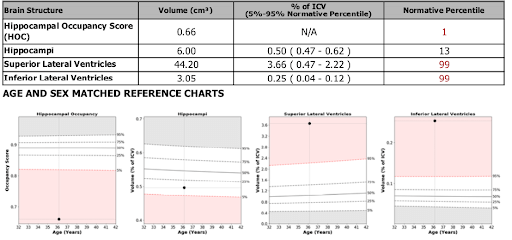
Case 2:

- A single T1 MRI scan of a 36-year-old female.
- HOC measures 0.66 cm3. The mesial temporal lobe is more than two standard deviations from the normative range for age and sex.
- The hippocampi demonstrate atrophy and are beyond two deviations below the normative range for age and sex.
- The lateral ventricles and inferior lateral ventricles are enlarged beyond two standard deviations of the normative range for age and sex.
Conclusion: Low hippocampal volume and suggestive of local ex-vacuo dilatation. This supports mesial temporal lobe-focused neurodegenerative etiology. Suggestive of the progression from MCI to AD.
This measure not only aids in the differentiation of individuals with congenitally small hippocampi from those with small hippocampi due to a degenerative disorder but also effectively demonstrates the risk of conversion from MCI to AD.
Citations:


