By
Cortechs.ai
2 mins
An integral component of Multiple Sclerosis (MS) management is the ability to proficiently assess disease activity on MRIs from new and enlarging T2 FLAIR lesions.
The latest release of the LesionQuant module of NeuroQuant (FDA cleared, CE marked) physicians can choose to also review of spatially aligned and reconstructed images of 3D T1 and FLAIR images along slide the FLAIR color overlay images. This additional feature offers a supplemental review of lesion segmentation provided by LesionQuant.
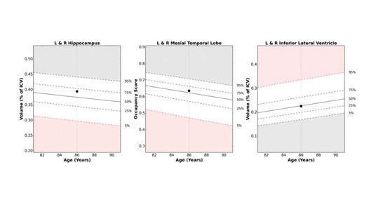
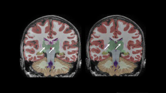
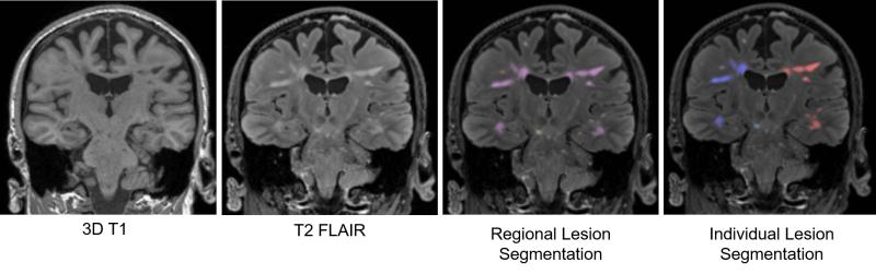 In addition, LesionQuant delivers fast, accurate anatomically color-coded segmented lesion images and automated FLAIR lesion and brain volume measurements compared to normative reference data. When a prior study is available, lesion change visualization and brain structure change data is available for review.
Want to learn more about LesionQuant?
We recently had a webinar to discuss the new features available in the LesionQuant from our latest software release. We also examined with case studies how the LesionQuant can help physicians quantify brain structure and lesions and as well as help in effectively evaluating disease activity in MS, based on current guidelines and research.
Our LesionQuant white paper provides more information about the accuracy and reproducibility available using LesionQuant for fully automated FLAIR lesion segmentation.
Share