In the evolving landscape of medical diagnostics, neuroimaging plays a critical role in understanding brain disorders, from Alzheimer’s to traumatic brain injuries. As AI-powered software advances and its algorithms become increasingly powerful, it holds the promise of significantly improving both the accuracy and efficiency of neuroimaging diagnostics.
The Challenges of Traditional Methods
While traditional diagnostic methods have been invaluable in the medical field, they come with several limitations. Human error, time-consuming processes, and the difficulty in detecting subtle changes in the brain over time can impact diagnostic outcomes. These challenges have highlighted the need for more efficient and consistent tools, such as AI-powered imaging solutions.
Alternate paragraph: Traditional volumetric analysis in the diagnostic workflow is often time-consuming and labor-intensive, requiring radiologists to manually segment brain structures from MRI scans. This process can take several hours per scan and is prone to variability, as it heavily relies on the radiologist’s expertise and experience. A study published in NeuroImage (DOI: 10.1016/j.neuroimage.2015.12.035) quantified this challenge, revealing that manual segmentation can take anywhere from 4 to 8 hours per scan, depending on the complexity of the brain structures involved. This extensive effort not only delays the diagnostic process but also introduces the risk of inconsistent measurements, making it difficult to accurately track disease progression. These challenges highlight the need for automated solutions like NeuroQuant, which significantly reduce analysis time and improve consistency.
The Benefits of AI-Powered Neuroimaging
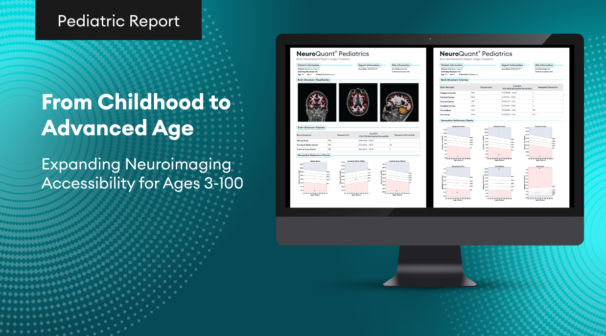
AI software like NeuroQuant significantly enhances the diagnostic workflow by providing automated, quantitative brain volume measurements, which streamline and improve the accuracy of diagnosing neurological conditions. Unlike traditional methods that rely on manual interpretation by radiologists—often leading to variability and longer processing times—NeuroQuant quickly analyzes MRI scans, offering objective data that supports faster and more consistent decision-making. For example, a study published in the American Journal of Neuroradiology (DOI: 10.3174/ajnr.A4509) demonstrated that NeuroQuant’s automated measurements were not only consistent but also correlated well with manual segmentation, proving its reliability and efficiency. This allows clinicians to detect brain atrophy and monitor disease progression more effectively, making it a valuable tool in routine clinical practice.
NeuroQuant demonstrates exceptional precision in brain volumetric analysis by providing consistent and accurate measurements of brain structures, which is crucial for diagnosing and tracking neurological conditions. Its automated software reduces the variability often seen with manual segmentation, leading to more reliable assessments of brain volume changes. For instance, a study published in Frontiers in Neurology (DOI: 10.3389/fneur.2019.01168) found that NeuroQuant’s measurements of hippocampal volumes were highly reproducible and closely matched manual expert assessments. This precision ensures that clinicians can detect subtle changes in brain atrophy over time, making NeuroQuant a valuable tool for monitoring disease progression in conditions such as Alzheimer’s disease, epilepsy, and multiple sclerosis..
Addressing the Limitations of AI
Despite its advantages, AI software also has its limitations. AI tools are only as effective as the data they are trained on, and robust data integration is critical to ensuring accurate results. Additionally, while AI can assist in diagnosis, the role of the physician remains crucial in interpreting the data and making final treatment decisions.
A key factor in overcoming these limitations is the quality and comprehensiveness of the datasets used to train the AI. In the case of NeuroQuant, its normative database provides a strong foundation for accurate and reliable results.
NeuroQuant’s normative database is built from thousands of age- and gender-matched brain scans, offering robust and clinically validated comparisons of individual brain structure volumes to a healthy population. By adjusting for key factors like intracranial volume, age, and gender, NeuroQuant ensures accurate and meaningful assessments of neurodegenerative conditions. With over 100,000 successfully processed MRI scans and sophisticated statistical models, the database provides fast, reliable, and clinically relevant volumetric insights that support confident diagnosis and treatment planning.
In conclusion, AI neuroimaging software like NeuroQuant offers powerful solutions that complement traditional diagnostic methods, enhancing both speed and accuracy. By incorporating AI, healthcare providers can leverage cutting-edge technology to deliver better outcomes for their patients.
Citations:
Aiello, M., Cavaliere, C., D’Albore, A., & Salvatore, M. (2019). The Challenges of Diagnostic Imaging in the Era of Big Data. Journal of clinical medicine, 8(3), 316. https://doi.org/10.3390/jcm8030316
Luo, W., Airriess, C., & Albright, J. (2023). The NeuroQuant® normative database: Comparing individual brain structures. Cortechs.ai.
