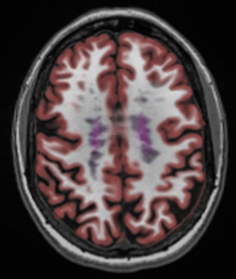
 Since MRI acquisition protocols used can have an impact on the reliability and accuracy of NeuroQuant’s MR image segmentation and volume measurements, the engineering and support teams at Cortechs.ai have put together a lot of great and pertinent information to help ensure our customers achieve the best possible outcomes when using Cortechs.ai products.
To optimize results, we advise the following:
1. Use the recommended scanner settings for each MRI manufacturers and models. Find them here.
2. Review the Frequently Asked Questions. Read those here.
3. Read the following blog posts:
To help make de-identifying data easy and painless, Cortechs.ai added a feature to CTXNode (our secure cloud processing system) allowing MRI scan data can be easily de-identified during the process of uploading to NeuroQuant. Read post.
Contrast agents are typically used to study vasculature, something that is not needed for volumetric MR imaging. NeuroQuant requires a non-contrasted, 3D T1 sagittal sequence to segment and measure brain structure volumes. Read post.
Alignment to atlas is an important step in NeuroQuant’s analysis process. To achieve its high quality segmentation results, NeuroQuant aligns each patient’s brain image to an atlas of the brain. Read post.
Patients who have shunts, implants or other artifacts or inherent abnormalities, can potentially generate segmentation misregistration. To prevent such errors and protect the accuracy of the results, NeuroQuant prevents analysis when such artifacts or anatomical anomalies are present. Read post.
3D T1 sagittal MRIs are needed to obtain ideal brain morphometry for volume analysis. In order to achieve its high quality segmentation results, NeuroQuant has been optimized for processing 3D T1 sagittal MRI acquisition sequence in 1.5 and 3.0T closed scanners. Read post.
If a sequence is acquired and uploaded to NeuroQuant with any of the X, Y or Z coordinates outside +/-100mm, automated volumetric analysis cannot be performed. An NeuroQuant error message will be returned. Read post.
4. Consult the NeuroQuant User Manual.
5. Contact the support team with any questions.
![]()
